Anatomy Rib Cage - Anatomy of the Rib Cage for Artists, Vidyaranya Arts - YouTube - Quads only geometries (no tris/ngons).. The bones of the rib cage are the sternum, the 12 thoracic vertebrae and the 12 pairs of ribs. Pain under the left rib cage can mean anything from a ruptured spleen, to heart trouble, to just needing to have a good fart. Related posts of rib cage diagram with organs woman stomach anatomy. Rib cage anatomy the rib cage, shaped in a mild cone shape and more flexible than most bone sets, is made up of varying elements such as the thoracic vertebra, 12 equally paired ribs, costal cartilage, and held together anteriorly by the sternum. Muscle anatomy drawing 12 photos of the muscle anatomy drawing anatomy muscle sketches, arm.
The ribs are a veritable collection of bone, muscle, and organs, most of which are fairly important for living and other useful functions. The upper edge is round and the lower sharp. Human rib cage anatomy 3d model. It consists of the 12 pairs of ribs with their costal cartilages and the sternum (figure 6.38). They run inferoanteriorly from the rib above to the rib below, and are continuous with the external oblique of the abdomen.
Rib Cage Anatomy Photograph by Collection Abecasis from images.fineartamerica.com At the chest, many rib bones connect to the sternum via costal cartilage,. On the interior wall of the rib body is a channel, sulcus costae, with blood vessels and nerves. It may occur after an obvious injury or without explanation. Anatomy the rib cage is a bony structure found in the chest (thoracic cavity). A rib has a flat body, as you can see from the picture of the anatomy of the human rib cage. As part of the bony thorax, the ribs protect the internal thoracic organs. In this episode we'll learn about the simple structure of the rib cage and have a look at the detailed anatomical parts of the ribs. Rib cage pain may be sharp, dull, or achy and felt at or below the chest or above the navel on either side.
4 individual objects (spine portion, ribs, cartilages, sternum), sharing the same non overlapping uv layout map, material and pbr textures set.
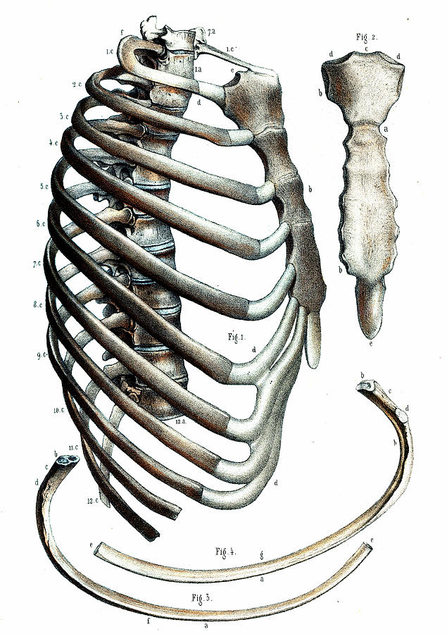
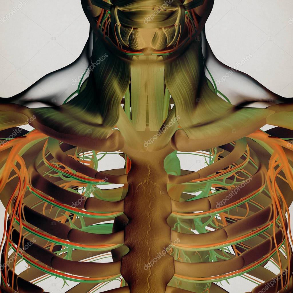
Originate at the lower border of the rib, inserting into the superior border of the rib below. At the chest, many rib bones connect to the sternum via costal cartilage,. Rib cage anatomy the rib cage, shaped in a mild cone shape and more flexible than most bone sets, is made up of varying elements such as the thoracic vertebra, 12 equally paired ribs, costal cartilage, and held together anteriorly by the sternum. Contributing to their role in protecting the internal thoracic organs. Quads only geometries (no tris/ngons). The thoracic cage consists of the 12 thoracic vertebrae, the associated intervertebral discs, 12 pairs of ribs with their costal cartilages, and the sternum. The sternum is a flat bone that is made up of three parts, the (1) manubrium, (2) body, and the (3) xiphoid process. Moreover, there are many vital organs such as the heart, liver, gall bladder, kidney, and lungs under your right rib cage. They run inferoanteriorly from the rib above to the rib below, and are continuous with the external oblique of the abdomen. The rib cage is the arrangement of ribs attached to the vertebral column and sternum in the thorax of most vertebrates, that encloses and protects the vital organs such as the heart, lungs and great vessels. Each pair is numbered based on their attachment to the sternum, a bony process at the front of the rib cage which serves as an anchor point. Woman stomach anatomy 7 photos of the woman stomach anatomy activate javascript anatomy of a woman body, female abdomen anatomy, female organ anatomy, human stomach anatomy, stomach anatomy and physiology, stomach anatomy antrum, stomach anatomy pictures, womens stomach anatomy, stomach, anatomy of a woman body, female. Human rib cage anatomy 3d model.
The ribs are curved, flat bones which form the majority of the thoracic cage. On the interior wall of the rib body is a channel, sulcus costae, with blood vessels and nerves. The lungs are two separate but connected organs located in the upper chest, covered by the rib cage. There are twelve pairs of ribs, all of which articulate with the vertebral column. Pain under the left rib cage can mean anything from a ruptured spleen, to heart trouble, to just needing to have a good fart.
Anatomy Rib Cage Picture - Human Anatomy Body from www.anatomylibrary99.com The top edge of the manubrium has a depression called the suprasternal or jugular notch. The purpose of the lungs is to take in oxygen from the environment and filter out any impurities or harmful pollutants. Rib cage anatomy the rib cage, shaped in a mild cone shape and more flexible than most bone sets, is made up of varying elements such as the thoracic vertebra, 12 equally paired ribs, costal cartilage, and held together anteriorly by the sternum. Each pair is numbered based on their attachment to the sternum, a bony process at the front of the rib cage which serves as an anchor point. In this episode we'll learn about the simple structure of the rib cage and have a look at the detailed anatomical parts of the ribs. The rib cage consists of 24 ribs, 12 on either side, and it shields the organs of the chest, including the heart and the lungs, from damage. The thoracic cage (rib cage) forms the thorax (chest) portion of the body. There are 11 pairs of external intercostal muscles.
This furrow isn't present in the 11th and 12th ribs.
Related posts of rib cage diagram with organs woman stomach anatomy. The cartilage strips are called costal cartilage (costal is the anatomical adjective that refers to the rib) and connect on their other end to the sternum. The rib below that is rib 2, and it connects to the t2 thoracic vertebra, and so on. In this video, we explore:1) the anatomy of the sternum2) the anatomy and differences between the three classes of ribs3) the anatomy and differences between. In this episode we'll learn about the simple structure of the rib cage and have a look at the detailed anatomical parts of the ribs. The ribs are a veritable collection of bone, muscle, and organs, most of which are fairly important for living and other useful functions. Related posts of muscle anatomy rib cage muscle anatomy drawing. At the chest, many rib bones connect to the sternum via costal cartilage,. Air reaches the lungs through the trachea, located beneath the throat. Originate at the lower border of the rib, inserting into the superior border of the rib below. However, only seven have a direct articulation with the sternum. The ribs are attached to the breastbone, which is the. As part of the bony thorax, the ribs protect the internal thoracic organs.
The cartilage strips are called costal cartilage (costal is the anatomical adjective that refers to the rib) and connect on their other end to the sternum. 4 individual objects (spine portion, ribs, cartilages, sternum), sharing the same non overlapping uv layout map, material and pbr textures set. Contributing to their role in protecting the internal thoracic organs. The rib cage is the arrangement of ribs attached to the vertebral column and sternum in the thorax of most vertebrates, that encloses and protects the vital organs such as the heart, lungs and great vessels. The lungs are two separate but connected organs located in the upper chest, covered by the rib cage.
Human rib cage anatomy model — Stock Photo ... from st3.depositphotos.com At the chest, many rib bones connect to the sternum via costal cartilage,. They articulate with the vertebral column posteriorly, and terminate anteriorly as cartilage (known as costal cartilage). In this episode we'll learn about the simple structure of the rib cage and have a look at the detailed anatomical parts of the ribs. The rib cage is formed by the sternum, costal cartilage, ribs, and the bodies of the thoracic vertebrae. Woman stomach anatomy 7 photos of the woman stomach anatomy activate javascript anatomy of a woman body, female abdomen anatomy, female organ anatomy, human stomach anatomy, stomach anatomy and physiology, stomach anatomy antrum, stomach anatomy pictures, womens stomach anatomy, stomach, anatomy of a woman body, female. The thoracic cage (rib cage) forms the thorax (chest) portion of the body. Originate at the lower border of the rib, inserting into the superior border of the rib below. A rib has a flat body, as you can see from the picture of the anatomy of the human rib cage.
Rib cage pain may start in one area but travel to an area nearby.
The bones of the rib cage are the sternum, the 12 thoracic vertebrae and the 12 pairs of ribs. The thoracic cage protects the heart and lungs. The ribs are a veritable collection of bone, muscle, and organs, most of which are fairly important for living and other useful functions. The lungs are two separate but connected organs located in the upper chest, covered by the rib cage. However, only seven have a direct articulation with the sternum. Rib cage, in vertebrate anatomy, basketlike skeletal structure that forms the chest, or thorax, and is made up of the ribs and their corresponding attachments to the sternum (breastbone) and the vertebral column. The thoracic cage (rib cage) is the skeletal framework of the thoracic wall, which encloses the thoracic cavity. The thoracic cage (rib cage) forms the thorax (chest) portion of the body. This furrow isn't present in the 11th and 12th ribs. Elevates the ribs, increasing the thoracic volume. Ten of the twelve ribs connect to strips of hyaline cartilage on the anterior side of the body. The ribs are curved, flat bones which form the majority of the thoracic cage. The thoracic cage consists of the 12 thoracic vertebrae, the associated intervertebral discs, 12 pairs of ribs with their costal cartilages, and the sternum.